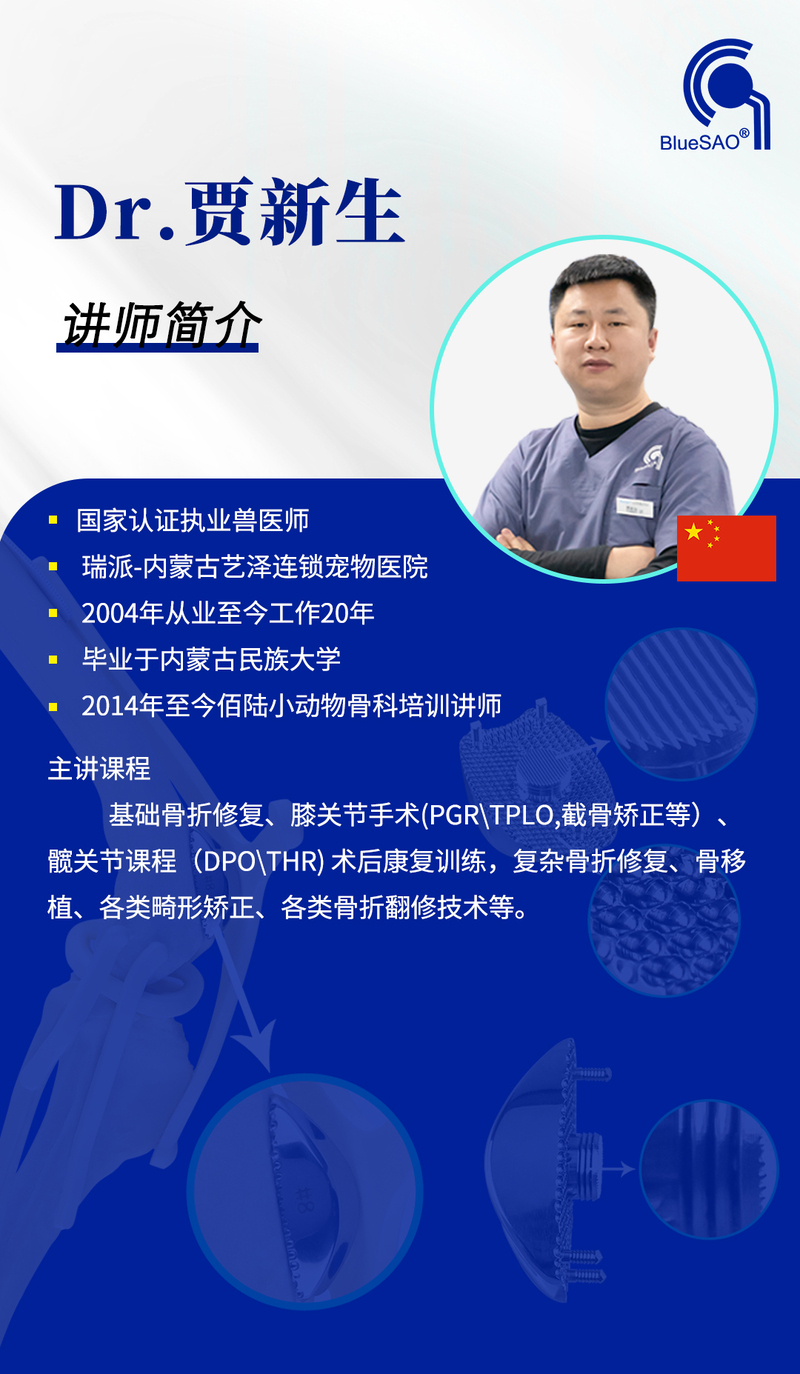
A New Perspective on the Treatment of Canine Patellar Luxation Proposed by Dr. Jia Xinsheng is Gaining Wider Acceptance Among Veterinarians.
A New Perspective on the Treatment of Canine Patellar Luxation Proposed by Dr. Jia Xinsheng is Gaining Wider Acceptance Among Veterinarians.
Dr.Jia’s viewpoint is: the Patellar Groove Replacement (PGR) can provide a pain-free joint for patellar luxation patients. He recommends PGR for eligible cases, such as those with a flat, deformed, or dysplastic trochlear groove, an excessively narrow groove, or cases where veterinarians assess that trochleoplasty (groove deepening) may not yield effective results.
Key Rationales:
1. **Trochleoplasty** is essentially an artificial reconstruction of the trochlear groove, and its degree of trauma is no less than that of PGR. Unskilled technique during trochleoplasty may lead to cartilage fragmentation, whereas PGR provides a wider, more anatomically conforming, and pain-free articular surface.
2. Trochleoplasty damages the articular cartilage, which cannot regenerate and is instead replaced by fibrocartilage. Fibrocartilage may undergo calcification and osteophyte formation.
3. Trochleoplasty cannot achieve the same width and depth as an artificial trochlear prosthesis, nor can it guarantee the prevention of future arthritis or ensure proper patellar tracking within the groove. Patellar reshapingand then forcing the patella into a narrowed groove may cause significant pain during movement.
The following case study effectively illustrates Dr. Jia Xinsheng’s perspective. A Yorkshire Terrier underwent **trochleoplasty on the left hindlimb** and **BlueSAO Press-Fit Biological PGR on the right hindlimb** on the same day. A noticeable difference in gait was observed just three days postoperatively.
Case Details:
- Patient: Tuesday, male Yorkshire Terrier, 3 years old, 3.67 kg
- History: Diagnosed with grade 2 medial patellar luxation at 7–8 months of age. One month prior, the owner observed occasional limping and sudden vocalization (approximately 5 episodes) during vigorous play, followed by right hindlimb lifting.
- Clinical Signs:
- Right hindlimb: Grade 3 medial patellar luxation upon palpation (without sedation).
- Left hindlimb: Grade 2 medial patellar luxation.
- Intraoperative Findings:
- Right hindlimb:** Defect above the medial trochlear ridge, dorsal patellar cartilage defect, and significant soft tissue hyperplasia laterally. **Press-Fit Biological PGR** was performed using a size 2 prosthesis.
- Left hindlimb: No severe trochlear defect observed; trochleoplasty + lateral tibial tuberosity transposition was performed.
- Surgical Plan:
- Left: Trochleoplasty + lateral tibial tuberosity transposition.
- Right: PGR + lateral tibial tuberosity transposition.
- Bilateral: Medial joint capsule release + lateral joint capsule imbrication.
- Postoperative Observations:
- The **right limb (PGR)** showed normal weight-bearing and smooth movement.
- The **left limb (trochleoplasty)** exhibited obvious pain and occasional toe-touching or lifting.
Recommendation:
For severe patellar luxation, trochlear hyperplasia, or significant trochlear wear, the surgical goal should be to reconstruct a lasting, pain-free joint with minimal trauma. In such cases, PGR is the optimal solution.

Orthopedic Instruments We Use BlueSAO